

Our covering for the first clean room required a “bunny suit” (a giant coverall, like little kid pjs), slip-on booties to cover our feet, goggles, a hair net, and nitrile gloves. However, this was a class 1000 clean room. There was a class 100 as well, which was even “cleaner”, meaning the controls on dust and other particles were even stricter. To our 1000-level outfit, we added higher booties that snapped shut at the knee, a hood, and a mask if we were worried about contaminating things with the saliva and other particles we exhaled. Here is a picture of me completely gowned in my full bunny suit and 100-level gear:
The clean rooms we were in are primarily used to create diffraction gratings and other optical gratings. These gratings change laser light into new and interesting patterns, and can even make holograms. To make a grating, we must first make a “mask”, or a basic template for the grating. This is basically like a mold for the grating, and s
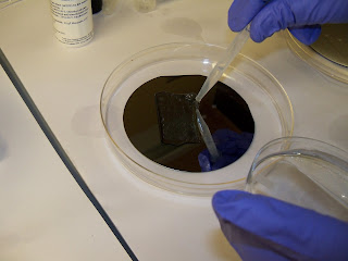
erves as our template. A laser is programmed by a computer to expose a chemical on a piece of chromium and photo-resist coated glass in a specific pattern. This chemicals can be washed off when it is exposed, leaving behind a pattern based on where the laser was. Here is another mask made by someone in the lab, you can see the chromium has been etched away, leaving clear glass in some places.

A lot of different chemicals are used in this process, such as sulfuric acid. Using these chemicals can be very dangerous, so sometimes more protection than the normal gloves and goggles is needed...

Our pattern was a series of equally spaced dots, about 50 micrometers across. (Can anyone convert this to meters? How about nanometers?) Next week we will take our mask and make the next part of our grating by using light to etch a pattern based on our mask.
(This post was originally made on 7/5/2010, but was reposted to a new blog for technical reasons)
It feels like this first week of BU's RET program has gone by so fast, we've gotten so little done! Here I will try and summarize the first week of working in the lab.
I am working with Ms. Sewell from Swampscott high school in the Biomedical Optics Laboratory. We were introduced to the lab space by the lab's head faculty member, Doctor Bigio, a former researcher at Los Alamos laboratories. Both Ms. Sewell and I have done research before, but in labs much more oriented towards chemistry, so seeing what is is like in an optics lab was quite a new experience! The benches were set up very differently, and most of the familiar glassware wasn't present. Instead, there are lasers and mirrors, designed to allow the researchers to use directed light in their experiments. Optics is a branch of physics and engineering concerned with the use of light. Many sensors that you are commonly familiar with, such as the scanner in a grocery store, use lasers and optics. Biophotonics uses these principles combined with biology to make new and interesting devices, such as a probe that can tell if the tissue it is touching is cancerous based entirely on how it reflects light.
Our project involves using the properties of light to determine mechanical properties of a single cell. Since the cells have a different refractive index than water (remember physics anyone?) any light shone through them will be bent. We can then detect that light and tell something about how dense the cell is.
This project takes it one step further. By hitting the cells with a burst of ultrasound, we can cause them to wiggle! This wiggle is seen by the light level at the place where we are detecting rising and falling. Theoretically, we can use this wiggle to determine how rigid the cells are.
Why is this important?
Healthy cells have a cytoskeleton, a framework holding them in shape. This makes them somewhat rigid, but with enough flexibility to function wherever they need to be in a body. It also helps the cell move. Cancer cells show a drastically different structure in the cytoskeleton, and we should be able to detect the resulting change in rigidity. Also, cells that are dying begin to lose their structure. They would get softer and oscillate (wiggle) differently.
Right now, the machine we are using doesn't quite work, but our job is to "optimize" it. This means get it to work, and to hopefully work very well. The first week was mostly about seeing the lab and getting to know where everything is. this next week we should really get down to finding out what will make this project take off!